Reflexes, Synapses, and Neurotransmitters

What are Reflexes, Synapses, and Neurotransmitters?

This page provides information about reflexes, synapses, and neurotransmitters. The explanations and examples are first. The free exercises are at the bottom of the page.

What are Reflex Actions?

Reflex actions are automatic responses within the body to external stimuli.

Reflexes are automatic in the sense that they are involuntary, and we don’t need to think about doing them.

Because we often use them in response to danger or potential injury, reflex actions are rapid responses.

What is a Reflex Arc?

The electrical impulses that are responsible for a reflex action travel along a pathway called a reflex arc.

The reflex arc pathway is as follows:

Receptors in the body transform stimuli into electrical impulses.

These nerve impulses go from the receptor and travel to a sensory neuron.

From the sensory neuron, the impulse goes to a relay neuron in the spinal cord. The impulse then goes to a motor neuron.

Finally, the impulse leaves the motor neuron and travels to an effector, where the reflex action is carried out.

What is a synapse?

A synapse is the place where two neurons join.

Remember that groups of neurons form nerves in the body.

Types of Synapses

Broadly speaking, synapses can be classified into two groups:

1) electrical synapses

2) chemical synapses

What are Electrical Synapses?

Electrical synapses pass ions and signaling molecules directly from one cell to another.

In other words, electrical synapses are a direct link between two cells in the body.

There is no synaptic delay in electrical synapses, so the response is very rapid.

What are Chemical Synapses?

No direct signal is passed between neurons in a chemical synapse.

Instead, there is a diffusion of the chemical molecules, also called neurotransmitters, in the synapse over time.

For this reason, the transmission of information via a chemical synapse is slower than that of an electrical synapse.

Remember that most synapses in the human body are chemical synapses.

How Do Chemical Synapses and Neurotransmitters Function?

When a nerve impulse travels to the end of one neuron, it creates a chemical. These chemicals are called neurotransmitters.

The chemicals are released into the gap between the neurons.

The chemicals then create an impulse in the next neuron.

This impulse continues along the reflex arc.

How Do Neurons and Synapses Work Together?

Synapses cause nerve impulses to be slowed down.

However, a neuron can form synapses with more than one neuron.

So, an impulse can pass to other neurons when it reaches the end of a sensory neuron.

Because synapses can be formed with several neurons at once, we are capable of responding to any given stimulus in more than one way.

For example, if you hear something unpleasant, you can cover your ears or walk away from the noise.

Synapses and Overriding Reflex Actions

In the spinal cord, a relay neuron forms synapses with a sensory neuron and with a motor neuron.

The spinal cord also forms synapses with neurons carrying nerve impulses from the brain.

These impulses from the brain can help you overrule a reflex action.

For instance, you may experience the reflex action of blinking when your picture is taken with a flash.

However, if you tell yourself to focus on a distant object instead of the flash, you may be able to override your reflex action and not blink.

Synapses and Neurotransmitters – Questions

1. Label the parts of a chemical synapse in the diagram below.

Reflexes, synapses, and neurotransmitters
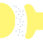
  • What do the two yellow areas represent?
  • What does the white gap in the middle represent?
  • What do the blue dots represent?

2. How is the signal transmitted in the illustration in question 1?

3. What is the most common type of synapse in the human body?

4.  A sensory neuron carries an impulse to a relay neuron in your spine, which passes it on to a motor neuron. How many synapses could be involved in this response?

5. You realize that you smell a fire in your house. Why is it advantageous that a neuron can form connections with many other neurons when you have to respond to a situation like this?

Synapses and Neurotransmitters – Answers

Answer 1
  • The two yellow areas represent neurons.
  • The white gap in the middle represents the synapse.
  • The blue dots represent neurotransmitters.
Answer 2

The diagram in question 1 represents a chemical synapse, so no direct information is passed in this synapse.

The signal is transmitted over time from the diffusion of the chemical molecules or neurotransmitters.

Answer 3

Most synapses in the human body are chemical synapses.

Answer 4

At least three synapses could be involved in the response.

In the spinal cord, a relay neuron forms synapses with both a sensory neuron and with a motor neuron. The spinal cord also forms synapses with neurons carrying nerve impulses from the brain.

Answer 5

The presence of multiple pathways is advantageous because you can decide what action to take first. The following are among the potential actions that you could take:

  • Scream “fire” to warn other members of your family
  • Walk to the kitchen to see if something is burning
  • Call the fire department
  • Run out of the house

More help with science

More science exercises

For exam downloads, please click on the image below.

More help with receptors and effectors

DNA

What is DNA?

DNA stands for deoxyribonucleic acid.

It contains the molecules that make up chromosomes and genes.

So, it contains the information that passes characteristics from one generation to another.

Spiral Structure

DNA is located in the nucleus of a cell.


It is made up of two interlocked spiral strands.

Genetic information is stored on the horizontal threads that connect the two spirals.

These threads store four different types of molecules in a code.

So, in the illustration below, the blue, pink, yellow, and  green segments of the threads are each different types of molecules.

DNA structure

Function and Instructions

The function of DNA is to embody genetic instructions.

You can think of it as being somewhat like the blueprint for a house.

Using the blueprint, the builder has to interpret the design and build the house.

In the same way, the cells have to interpret and translate the DNA into action in the cells in the body.

DNA tells the cells what proteins to make and gives the protein its particular individuality.

It also communicates with the cytoplasm in the cells.

To understand the role of cytoplasm, you may want to view our post on cell structure.

Mutations

The most important point to remember about DNA mutations for your exam is that mutations normally occur very infrequently.

If DNA fails to copy itself correctly during reproduction, mutations can occur.

Mutations can be deadly because they change the way that certain cells work.

Mutations that occur in germ cells can cause variations to the offspring of an organism.

Mutations can also be caused by environmental factors.

DNA in Cell Replication

DNA replication takes place before mitosis can occur.

Just before cell division takes place, each part of the pair of chromosomes contain a double helix of DNA, which originates from the parent cell.

This is copied so that it can be passed from cell to cell during cell division.

Because of mitosis, the replicated cells have the same DNA as the parent cells.

DNA can reproduce itself by spitting in half, in much the same way as a zipper unzips.


Each thread can copy the missing thread to regenerate the genetic code.

Other Nucleic Acids

Nucleic acids are synthesized in chemical reactions controlled by enzymes.

Nucleic acids are long chains of smaller units called nucleotides.

RNA is another type of nucleic acid.

RNA is a single stranded nucleic acid.

Chromosomes, Genes, Proteins, and RNA

You may need to compare and contrast chromosomes, genes, proteins, and RNA for your exam.

A gene is a distinct sequence of nucleotides that form part of a chromosome.

A chromosome has  a long coiled  strand of DNA in its nucleus, with some special proteins and some RNA.

The main role of RNA is to act as a messenger to carry instructions from DNA.

Exercises

  1. What is the structure of DNA?
  2. What is the function of DNA?
  3. What happens when mutations occur in germ cells?
  4. What are nucleic acids?
  5. What is RNA?

Answers

  1. It is made up of two joined spiral strands. There are horizontal threads that connect the two spirals. These threads store the genetic code.
  2. It tells the cells what individual proteins to make and communicates with the cytoplasm in the cells.
  3. These mutations can result in variations to the offspring of an organism.
  4. Nucleic acids are chains of nucleotides.
  5. RNA acts as a messenger to carry instructions from DNA.

Now look at our post on cell differentiation.


Meiosis

Meiosis Definition

In meiosis, the number of chromosomes in a cell is halved.

The daughter cells receive only one chromosome from each pair of chromosomes.

So, chromosomes, which occur naturally in pairs, are separated from their other half during meiosis.


Meiosis – The Essential Principal

The essential principal of meiosis is that it consists of two divisions of the cell nucleus, resulting in four cells that contain only one individual chromosome.

Unlike mitosis, the new cells do not contain pairs of chromosomes.

So, after meiosis is complete, each cell consists of only half of the number of chromosomes of the original cell.

Meiosis and Fertilization

Meiosis occurs in the formation of sex cells.

These sex cells are called gametes.

A gamete is a cell that cannot develop any further unless it unites with another cell.

Eggs and sperm are two examples of gametes

During fertilization, the nuclei of the gametes combine.

If gametes were formed by mitosis instead of meiosis, the amount of chromosomes would double with each succeeding generation.

Meiosis insures that the correct amount of chromosomes are maintained during the fertilization process.

Meiosis I and II

Meiosis actually consists of two successive divisions.

First of all, the parent cell splits in two. During the first meiotic division, the chromosomes line up together.

After the chromosomes line up, they separate and go to different cells.

During the second meiotic division, the products of the first division split into two again.

So, after the second meiotic division, there are a total of four daughter cells.

Each of the daughter cells is genetically different.

No DNA replication occurs between the first and second meiotic division.

Therefore, meiotic division encourages genetic variation.

Haploid Cells

After meiotic division, each daughter cell is a haploid cell.

As opposed to the diploid cells that are formed in mitosis, the chromosomes in haploid cells do not have a partner of the same identical length and of the exact same genetic material.

In other words, a haploid cell has only one chromosome.

This single chromosome is half of the original chromosome pair.

So, each chromosome is unique in a haploid cell.

The Eight Stages

The stages of meiotic division are similar to those of mitosis.

Prophase I – The chromosomes become elongated strands before the chromosomes begin to pair.

Metaphase I – The envelope that contains the nucleus disappears and the chromosomes begin to line up at the equator of the cell.

Anaphase I – The chromosomes move towards the opposite ends of the cell.

Telophase I – Chromosome movement is complete. Two new cells are formed.

Prophase II – The chromosomes, which consist of members of each pair, condense.

Metaphase II – Chromosome align in the center of each spindle.

Anaphase II – The two chromatids of each pair move to opposite ends of the cells.

Telophase II – Four haploid cells are formed, each containing a haploid nucleus.

Only one of these four products develops into a functional cell.

The other three cells disintegrate.

The cell that survives contains only half of the pair of chromosomes from the original cell.


The complete process of meiotic division can take several days or even weeks.

Compare and Contrast Meiosis and Mitosis

Similarities

Both processes refer to cell division.

Both processes have the stages of prophase, metaphase, anaphase, and telophase.

Differences

Mitosis usually lasts about an hour, while meiosis can last for days or even weeks.

In meiosis, the amount of chromosomes halves, while in mitosis, the amount of chromosomes is maintained.

Meiosis I and II

Exercises

  1. What are gametes?
  2. What is the essential principal of meiosis?
  3. What is a haploid cell?
  4. How are meiosis and mitosis similar?
  5. How are meiosis and mitosis different?

Answers

  1. A gamete cell cannot develop without uniting with another cell.
  2. After meiosis, each cell contains just half of the number of chromosomes of the original cell.
  3. In haploid cells, each chromosome is unique. The chromosomes in haploid cells do not have an identical partner.
  4. Both processes refer to cell division and have the same stages (prophase, metaphase, anaphase, and telophase).
  5. In meiotic division, the amount of chromosomes halves, while in mitotic division, the amount of chromosomes is maintained.

You may also want to look at our posts on cellular organelles and cellular differentiation.


Mitosis

Mitosis Definition

Mitosis refers to one of the processes of cell division.

Mitosis involves chromosomes, which occur naturally in pairs and which contain the organism’s genetic information.

In mitosis, chromosome pairs separate and then form new pairs.

Mitosis ensures that the new cell (called the daughter cell) has the same chromosome number as its parent cell.


So, genetic information is shared equally among new cells when mitosis occurs in complex organisms.

Mitosis – The Essential Principal

The most important point about mitosis is that the daughter cells receive exactly the same number and types of chromosomes as the parent cell.

So, the genetic constitution of the organism is maintained.

The Process of Mitosis – Step by Step

The process of mitosis consists of four basic steps:

  1. At the beginning of the division of the cell nucleus, each chromosome exists as a replicated structure. So, the DNA of the parent cell has already replicated before mitosis begins.
  2. Each chromosome then divides longitudinally (length-wise), forming two identical halves that separate from each other.
  3. The separated halves then move in opposite directions.
  4. Each of the chromosome halves becomes incorporated into the nucleus of one of the two new daughter cells that are formed.

Mitosis – The Scientific Stages

Scientifically speaking, mitosis consists of four stages.

The names of these four stages are:

  1. prophase
  2. metaphase
  3. anaphase
  4. telophase

The entire process of mitosis usually takes about an hour.

Prophase

Chromosomes condense and form distinct threads within the nucleus of the cell.

The chromosomes are already longitudinally double, meaning that they contain two identical halves.

Mitosis prophase

Metaphase

The chromosomes move to the center of the cell.

The chromosomes then form a line in the center of the cell.

This line is exactly halfway from each of the two ends of the cell.

Mitosis metaphase

Anaphase

During this phase, the two parts of each chromosome begin to divide and move to opposite ends of the cell.

Each of these two parts of the chromosome is call a chromatid.

When the separation is complete, each chromatid becomes a new chromosome.

At the end of anaphase, there will be two identical groups of chromosomes at each end of the cell.

Mitosis anaphase

Telophase

During telophase, an envelope or sac forms around each group of chromosomes.

Each half of the cell forms a new nucleus, and the chromosomes condense and become less spindle-like.

This means that, when viewed under a microscope, the chromosomes will have changed in appearance.


At the end of telophase, the cell divides into two new cells.

Mitosis telophase

Mitosis and Meiosis

Mitosis should not be confused with meiosis.

Meiosis halves the number of chromosomes, while mitosis maintains the number of chromosomes.

Exercises

  1. What is the basic principal of mitosis?
  2. What occurs during prophase?
  3. What occurs during metaphase?
  4. What occurs during anaphase?
  5. What occurs during telophase?

Answers

  1. The basic principal of mitosis is that daughter cells receive exactly the same number and types of chromosomes as the parent cell.
  2.  During prophase, chromosomes form distinct threads within the nucleus of the cell.
  3. During metaphase, chromosomes form a line at the exact, equidistant center of the cell.
  4. During anaphase, the two parts of each chromosome (called chromatids) begin to divide and move to opposite ends of the cell, forming two identical groups of chromosomes at each end of the cell.
  5. During telophase, an envelope forms around each group of chromosomes. Each half of the cell forms a new nucleus. The chromosomes condense and the cell divides into two new cells.

You may also wish to visit our posts on cell differentiation, cell structure, and meiosis.

Cell Differentiation

What is Cell Differentiation?

Cell differentiation occurs when a cell changes into another type of cell or cells.

The process of cell differentiation occurs during cell growth.

Cellular differentiation occurs in multicellular organisms.


Cells differentiate when they change from a zygote into a more complex cellular system.

A zygote is a single-celled eukaryotic cell, formed when reproductive cells unite at fertilization.

The Process of Cell Differentiation

When cells have fully differentiated, they become specialized.

For this reason, cell differentiation is sometimes referred to as cell specialization.

After cell differentiation:

  1. Specialized cells undergo chemical changes in their cytoplasm.
  2. Specialized cells take on unique shapes.
  3. Cells then do one specialized job.

The changes in the shapes of specialized cells, as well as the chemical changes, enable the cells to perform their unique jobs within the organism.

In this way, cells are adapted to their special function to become well suited to the jobs they have to do.

Examples of Differentiated Cells in Plants

The following list provides two examples of differentiated cells in plants, as well as details about their shapes and functions.

Palisade Cell – Found on the top side of the leaf. Full of chloroplasts, which help with the process of photosynthesis.

cell differentiation 1

Root Hair Cell – Root hair cells are long and thin. They increase the surface area of the root of the plant, helping with the absorption of water and minerals.

cell differentiation 2

Examples of Differentiated Cells in Animals


The following list shows four examples of differentiated cells in animals.

After the name of each differentiated cell, you will see details about its shape and function.

Sperm Cell – The cell has a tail, allowing it to move freely in order to fertilize the ovum.

Ovum Cell – The ovum cell is larger than the sperm. The ovum has no active movement. This allows it to await fertilization by the sperm.

Ciliated Epithelial Cell – Cilia are small hair-like structures that are found in the respiratory system. They help to filter the air and to move mucus.

Nerve Cells – Nerve cells are made up of long fibers. Impulses travel along the fibers to the brain.

Cell Differentiation in Embryonic Stem Cells

Embryonic stem cells can change into any type of cell.

Before differentiation, the cells in the embryo are all the same. In other words, the cells are undifferentiated.

A fertilized egg divides during the process of mitosis, producing a group of new cells that form the embryo of the new organism.

Stem cells divide to produce more stem cells or to create specialized cells, such as blood cells.

Cells, Tissues, and Organs

Tissues are made when a group of similar cells come together.

A group of tissues come together to make an organ.

A group of organs come together to make an organ system or complete organism.

Cell Differentiation – Exercises

After you have read the material above, try to commit the key points to memory.

Then answer the questions that follow.

When you have finished, check your answers in the next section.

  1. What is a zygote?
  2. What three phenomena occur in cells after differentiation?
  3. Give two examples of differentiated cells in plants.
  4. Give two examples of differentiated cells in animals.
  5. “The shape of a specialized cell aids its function.” Give an example of a specialized cell and explain this phenomena.

Cell Differentiation – Answers

  1. A zygote is a single-celled fertilized cell.
  2. After cell differentiation, cells undergo chemical changes, take on unique shapes, and perform specialized jobs.
  3. The palisade cell and the root hair cell are two examples of differentiated cells in plants.
  4. The sperm cell, ovum cell, ciliated epithelial cell, and nerve cells are examples of differentiated cells in animals.
  5. The sperm cell is one example of a specialized cell. The shape of the tail on the sperm cell enables it to move freely in order to fertilize the ovum.

For more information on cell differentiation, please visit Biology Online.

You might also want to view our posts on cellular organelles, mitosis, and meiosis.

Cellular Organelles

What are Cellular Organelles?

All cells contain cytoplasm, which is the substance on the inside of the cell.

Organelles are the structures within the cytoplasm.

Organelles consist of many different structures.

What are the Functions of Organelles?

Organelles relate to the cell as the organs relate to the human body.


For instance, some organelles have a digestive system which helps to break down complex molecules within the cell.

Of course, organelles have a completely different structure than bodily organs.

Classes of Organelles

Two classes of organelles are called mitochondria and plastids.

Mitochondria

Mitochondria are present in almost all types of eukaryotic cells.

From our post on cell structure, you will remember that eukaryotic cells contain a nucleus.

So, eukaryotic cells include both plant and animal cells.

The mitochondria in eukaryotic cells produce energy, oxidize glucose substances, and release adenosine triphosphate (also known as ATP).

Under an electron microscope, mitochondria appear as small rods.

Cells that need to expend large amounts of energy can contain unusually large numbers of mitochondria.

Plastids

Plastids are the granules and incisions found in the cytoplasm of the cell.

They are found in plant cells.

Plastids may be oval or spherical in shape.

The two main kinds of plastids are amyloplasts and chloroplasts.

Amyloplasts are the starchy grains in plants in which carbohydrate is stored.

Chloroplasts contain the green pigment chlorophyll, which is essential in the process of photosynthesis.

Organelles in Eukaryotic Cells

In addition to mitochondria and chloroplasts, eukaryotic cells can also contain the following organelles.

Endoplasmic Reticulum

There are two types of endoplasmic reticulum (also known as ER): rough endoplasmic reticulum and smooth endoplasmic reticulum.

Rough endoplasmic reticulum is studded with ribosomes and is responsible for the isolation and transportation of the proteins that have been synthesized by the ribosomes.

Smooth endoplasmic reticulum does not contain ribosomes. Smooth endoplasmic reticulum is responsible for the expression of lipids.

Both types of endoplasmic reticulum form channels through the cytoplasm and therefore help to distribute substances more quickly around the cells.

Flagellum

The flagellum brings locomotion and sensory aspects to the cell.

Many organisms move by means of flagella.

Golgi Apparatus

The Golgi apparatus is an especially dense part of the cytoplasm.

As proteins leave the ER, the move to the Golgi apparatus, where carbohydrate is added.

Thus, the functions of the Golgi apparatus are the sorting, packaging, processing, and modification of proteins.


So, protein is packaged and processed by the Golgi apparatus before it leaves the cell.

Questions on Organelle Function

  1. What are the two classes of organelles?
  2. What is the function of mitochondria?
  3. What is the difference between rough endoplasmic reticulum and smooth endoplasmic reticulum?
  4. What is the primary function of the flagellum?
  5. What are the functions of the Golgi apparatus?

Answers to Questions on Organelle Function

  1. The two classes of organelles are called mitochondria and plastids.
  2. Mitochondria produce energy, oxidize glucose substances, and release adenosine triphosphate.
  3. Rough endoplasmic reticulum is responsible for the transportation of the proteins that have been synthesized by the ribosomes, while smooth endoplasmic reticulum is responsible for the expression of lipids. Rough endoplasmic reticulum contains ribosomes, but smooth endoplasmic reticulum does not.
  4. The flagellum is responsible for locomotion or movement.
  5. The functions of the Golgi apparatus are the sorting, packaging, processing, and modification of proteins.

Other Organelles

The vacuole and the nucleus are also organelles.

The vacuole is essential for storage and transportation, and helps maintain homeostasis.

The nucleus supports DNA maintenance, controls all activities of the cell, and is crucial for RNA transcription.

For more information on the vacuole and nucleus, please see our post on cell structure.

Cell Structure

Cell Structure and Organisms

Students who are going to study in health or science-related fields will need to understand cell structure for their college entrance exams.

This post will have a look at cell structure, including single-celled organisms like bacteria and yeast.

It will also discuss the structure of plant cells and animal cells.


When discussing cell structure, it is important to remember that cells are the building blocks from which all living organisms are made.

Single-Celled Organisms

Bacteria and other single-celled organisms can be identified as such because these cells do not have a nucleus.

Because of this, single-celled organisms are also called prokaryotic cells.

(“Pro” means “before” and “karyon” means “nucleus.”)

Bacterial cells are in capsule form. Their structure consists of a cell wall on the exterior and a substance called cytoplasm on the interior.

The cell wall provides strength to the cell and helps to prevent the cell from bursting.

Animal and Plant Cells

Animal and plant cells are more complex organisms. Each of these types of cells contain a nucleus.

Animal and plant cells are called eukaryotic cells.

(“Eu” means “true” and “karyon” means “nucleus.”)

Animal cells consist of an exterior plasma membrane, and cytoplasm and a nucleus on the interior.

Plant cell structure is perhaps even more complex.

Plant cells consist of an exterior cell wall which encases a plasma membrane.

Within this, there is cytoplasm, which contains chloroplasts.

In the center, plant cells contain a space called a vacuole. Therefore, the nucleus of a plant cell is slightly off-center.

Cell structure and the plasma membrane

The plasma membrane acts as a barrier between the cell and its environment.

This membrane also controls the movement of substances into and out of the cell.

Cell structure and cytoplasm

The cytoplasm performs most of the essential “operational” functions of the cell.


Cytoplasm is present in bacterial cells, animal cells, and plant cells.

Cell Structure – Review Questions

If you are taking an exam such as the HOBET or Nursing School Entrance Exam, you should be sure that you can easily answer the following questions on cell structure.

  1. Which cells are prokayyotic?
  2. Which cells are eukaryotic?
  3. How are plant and animal cells similar to bacterial cells?
  4. How are plant and animal cells different than bacterial cells?
  5. How are plant cells different than animal cells?

Answers to Cell Structure Review Questions

  1. Bacterial cells are prokaryotic, meaning that they do not have a nucleus.
  2. Animal and plant cells are eukaryotic. They have a nucleus.
  3. All of these cells contain cytoplasm.
  4. Plant and animal cells contain a nucleus, but bacterial cells do not have a nucleus.
  5. Plant cells contain vacuoles and chloroplasts, but animal cells do not.